
INDIA IMAGING - Advanced Diagnostic Center
As a medical student, it was my vision to assimilate world class technology into healthcare delivery in my city Deoghar. Many bits and pieces of that dream have been achieved in the last 1 year and the endeavour continues.
This website serves two purposes. One of these is a continuing education program for me and my colleagues in the medical profession, in our quest for the best answers to health problems. The focus here is on imaging technology . The other purpose is to provide information on CT Scan, ultrasound, X ray to patients and provide a guide map of facilities available at The India Imaging Advanced Diagnostic Center, Deoghar (Jharkhand), where I work.

About me
Dr. Bikram Mohan commenced his medical studies at the Rajendra Institute of Medical Sciences, Ranchi, Jharkhand in 1999, and, after completing his MBBS went on to do a post graduate degree in Radiology which he completed in 2011.
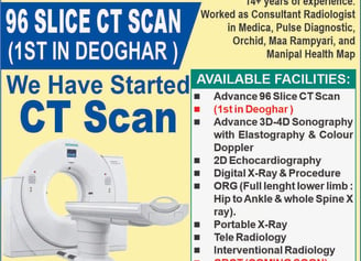
He has 14+ years of experience and worked in all premium hospitas of ranchi as Consultant Radiologist (Rajendra Institute of Medical Sciences, Bhagwan Mahaveer Medica Hospital, Pulse Diagnostic Center, Orchid Hospital, Maa Rampyari Hospital, Manipal Health Map etc). He has worked with all leading teleradiology company in India. Presently working as consultant teleradiologist with Manipal HelathMAP.
Armed with this experience he went on to establish a private diagnostic facility in Deoghar, with the vision of providing world class radiology services.
He is a member of several academic societies. (IMA, IRIA, ISA)

Recent Posts
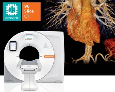
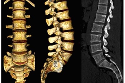
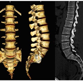
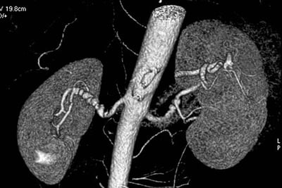
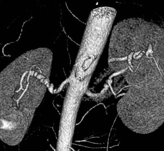
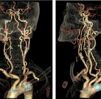
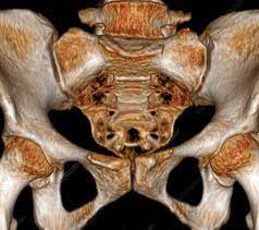
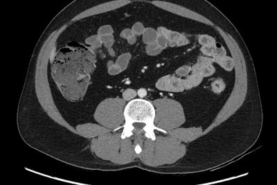
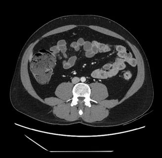
About 96 Slice CT SCAN
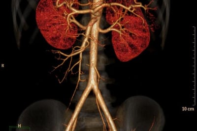
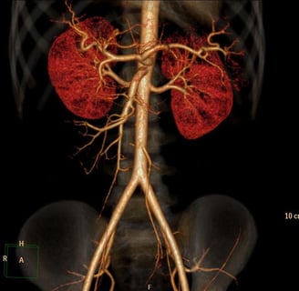
(Non-invasive CT Angiography)
Breakthrough technology and better results
India Imaging is the first to introduce 96 Slice CT scanner. The advanced scanning technologies, combined with our CT specialist, are able to provide the highest level of accuracy for diagnosing all types of illnesses in the body. These equipment are capable of performing cutting-edge tests like Pulmonary Angiograms, Brain Angiograms, True-match imaging, etc.
We are the first to introduce 96 Slice CT into the city. The state-of-the-art 96 slice CTs provide ultra-fast volume imaging. Further, all routine CT scans are done using ultra-thin slices with volume acquisition and isometric reconstruction that can be obtained in any plane. 3D-CT examinations are routinely performed along with a virtual colonoscopy, virtual bronchoscopy, and whole-body scanning. Patients pay the same charges for routine CT scans as they would at any other facility that does not have these high-quality machines.
Non-invasive imaging technologies continue to revolutionize every subspecialty of medicine.
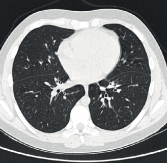
This scanner has a special x-ray tube and rotation speed, making it capable of performing very rapid scanning. It is used for performing non-invasive CT angiograms of the heart, brain, and other blood vessels of the body. The 96 slice configuration also provides breakthrough performance in advanced pulmonary imaging, multi-organ trauma evaluation, and low-dose paediatric applications to boost clinical capabilities to the highest level attainable.
96 Slice CT Scanner
India Imaging's latest 96 Slice CT provides the benefits of high-resolution, low-dose scanning with increased integration and collaboration. It provides consistent image quality across a diverse patient population and a wide range of exam types, enabling healthcare organizations to increase their care capabilities to treat more patients.
This high-end CT enables us to do scans in one go of the neck, chest and abdomen. Also, we can do triple-phase studies of the abdomen with sub-millimetre scan thickness. This state-of-the-art CT machine is capable of doing high-resolution CT Angio of the abdomen down to the feet in one acquisition and at a lower contrast dose. Other applications include brain perfusion, lung nodule analysis, and vessel analysis, allowing you to care for a wide range of patients with ease and efficiency.
The contrast in an image is the most important part in differentiating between a lesion and a normal parenchyma or a normal structure. The latest technology embedded enhances the differentiation between a lesion and normal parenchyma, it helps identify lesions that are very iso-dense or iso-attenuating which would otherwise be difficult to differentiate from normal parenchyma. Multiple advanced applications on the console allow for post-processing without a stand-alone workstation.
Features:
Enhance diagnostic confidence with superb image quality
See more detail across a range of patient types
Provide exceptional image quality for even small patients
Streamline the workflow and reduce variability
Provide exceptional image quality for even small patients
The 70 kV scan mode – the first in a system of its kind – helps take patient care to a new level by offering low-dose scanning of smaller patients and allowing for protection of radiation-sensitive organs. This scan mode offers up to 20% lower-dose scanning than 80 kV.
Enhance diagnostic confidence with superb image quality
The award-winning technology found in our CT systems is one of the ways that CT is able to offer such excellent and exceptional image quality at low doses. It helps personalize image quality based on patient needs at a low dose and improves image quality through artefact prevention and increased spatial resolution at a low dose. It also allows for improved tube efficiency.
your text here...


“This space is perfect for your favorite quote that inspires you.”
- Name Surname
Get our weekly newsletter
Sign up for our newsletter and never miss the newest blog post.